Regain Full Knee Function with World-Class ACL Reconstruction in India
Does knee pain or instability make it hard for you to enjoy the activities you love? Whether it’s playing sports, climbing stairs, or even walking comfortably, an ACL injury can disrupt your life in ways you never imagined.
Ignoring an ACL tear won’t make it go away—it can worsen over time, leading to chronic pain and even permanent joint damage. These injuries not only affect your mobility but also your confidence and independence.
With advanced ACL reconstruction in India, you can regain full knee function and return to the activities you love. Backed by top orthopedic surgeons, cutting-edge technology, and personalized care, ACL reconstruction gives you the confidence to move pain-free and reclaim your active life.
Let’s explore how you can take the first step toward recovery!


Swapnil’s Journey to Recovery
Let’s understand Swapnil’s experience with ACL reconstruction in India. Swapnil is an IT professional working in Mumbai, Maharastra, India.
He was a 29 years old bachelor, four years ago, and was living with friends in a flat. He used to go to the office on his newly purchased bike along with one of his friends Manish.
Swapnil was a jolly guy and loved to visit new places whenever he used to get time. He also used to go for tracking, every weekend. Like any other bachelor’s in Mumbai, he never missed an opportunity to go to a party with his friends.
One regular day, while he was going back home from the office he met with an accident. A car driver lost control of the vehicle while reversing his car from parking. Suddenly the car came on mid-road. Swapnil and his friend were on the same road. The car hit the moving bike pretty badly.
Swapnil and his friend fell down on the road. Due to a sudden fall, both of them were injured severely. Knee was the first body part to hit the road and both of them got bruises on their knees. The cut of Swapnil was so deep that even bone was visible.
Nearby people rushed to help and helped Swapnil and Manish to stand and supported them to sit in the car. The driver of the car was courteous enough to take them to the hospital. On the way to the hospital, Swapnil’s knee got swollen.
The doctor and nurse cleaned the wound and while cleaning the doctor could see some plastic parts and a small road stone in the knee. After cleaning the wound thoroughly, the doctor used switchers to close the wound. There were 16 stitches on Swapnil’s knee and 8 stitches on Manish’s knee.
In the meantime, an orthopedic doctor came and recommended the XRay to check the fracture. No fracture or dislocation is found in the Xrays report.
Looking at the swelling, the doctor suggested an MRI scan to check the ligament and condition of the knee.
In MRI, an ACL tear was clearly detected in Swapnil’s report.
The doctor described the condition of the knee and recommended surgery to fix the ACL.
Swapnil’s friend who was working in MedicoExperts got to know about this and he connected Swapnil with the best orthopedic surgeon in Mumbai. Swapnil was worried about his injuries and their impact. Even he started thinking about whether he would be able to walk normally. What will the recovery time be and will there be a big scar from the surgery on his knees?
After having the video consultation with the doctor and seeing all the reports, the doctor explained the procedure to Swapnil and addressed his concerns with respect to the long-term repercussions of the injury.
Swapnil got fully satisfied and went for the arthroscopic ACL reconstruction. Surgery was conducted in less than an hour with the hairline cut.
Swapnil was discharged from the hospital in a few hours when he recovered from anesthesia.
After discharge, the doctor advised Swapnil to change the dressing twice a week, put ice packs daily, knee elevation while sleeping, and the amount of load to give on feet. The doctor also recommended physiotherapy once the wound of the surgery is healed up.
With perfect surgery and nice care, Swapnil was able to walk with any support within a fortnight.
Let’s understand what is ACL tear is.

What is the Anterior Cruciate Ligament (ACL)?
The knee is a hinge joint with four ligaments that hold it together. A ligament is a structure in the knee that helps to control joint movement or motion by holding the bones together. There is a ligament on each side of the knee (the collateral ligaments) and two ligaments deep inside the knee.
The anterior cruciate ligament (ACL) and the posterior cruciate ligament (PCL) are two ligaments inside the knee that “cross” each other (PCL). Both ligaments attach to the top of the shinbone on one side and the end of the thighbone (femur) on the other (tibia).
One of the most important ligaments that helps stabilize your knee joint is the anterior cruciate ligament (ACL). The ACL is a ligament that connects your thighbone (femur) to your shinbone (tibia).


What are ACL Injuries and Common Causes?
A tear or sprain of the anterior cruciate ligament (ACL) — one of the strong bands of tissue that connects your thigh bone (femur) to your shinbone (tibia) — is known as an ACL injury.
ACL injuries often occur during high-impact sports or activities involving sudden stops, jumps, or direction changes. Common causes include:
- Sudden pivots or twists during sports.
- Direct impact during contact sports like football or basketball.
- Missteps or accidental falls.
It’s possible that your knee will swell, become unstable, and become too painful to bear weight on.
What are some signs and symptoms of an ACL injury?
The following are common signs and symptoms of an ACL injury:

What are the different types of ACL injury or tears?
ACLs that have been mildly damaged, such as when the ACL is mildly stretched but still provides adequate stability to the knee joint.
ACL injuries are uncommon and refer to a stretched and partially torn ACL.
When the ACL is completely torn in half and no longer provides any stability to the knee joint, it is classified as a grade 3 ACL tear.
Diagnosis of ACL Injuries
To rule out a bone fracture, X-rays may be required. Soft tissues, such as ligaments and tendons, are not visible on X-rays.
An MRI creates images of both hard and soft tissues in your body using radio waves and a strong magnetic field. An MRI can reveal the extent of an ACL injury as well as signs of damage to other knee tissues, such as the cartilage.
Ultrasound can be used to check for injuries in the ligaments, tendons, and muscles of the knee by using sound waves to visualize internal structures.
1. Bracing
This conventional technique is the most appropriate procedure for people who need correction of moderate to significant signs of aging. In this, incisions are sited around the ears, into the hairline, and also a small incision below the chin. This procedure yields the most dramatic and long-lasting results for most of the patients.
2. Physiotherapy
Conservative Management is often combined with the intensive rehabilitation program to regain the normal range of motion, increase stability and strength at the joint so as to perform activities of daily living.
3. Conservative management
This is indicated in cases of Acute and Partial ACL tear, in which proper rest is advisable with limited to no mobility in order to protect the joint and heal the ligament.
1. ACL Repair Surgery
In this procedure, the partially or completely torn ACL ligament is repaired by sewing it back together using sutures. However, studies have shown that the ACL repair generally fails over time and may require revision surgery or reconstruction.
2. ACL Reconstruction Surgery
ACL reconstruction is surgery to replace a torn anterior cruciate (KROO-she-ate) ligament (ACL) — a major ligament in your knee.
Ligaments are strong bands of tissue that attach one bone to another bone. During ACL reconstruction, the torn ligament is removed and replaced with a band of tissue that usually connects muscle to bone (tendon). The graft tendon is taken from another part of your knee or from a deceased donor.
In this procedure, the torn ACL ligament is replaced by using a substitute graft taken from either of the following sources
- Hamstrings Muscle Tendon AutoGraft taken from the patient’s own knee
- Patellar Tendon AutoGraft is taken from the patient’s own knee
- Quadriceps Muscle Tendon AutoGraft
- Allograft is a graft taken from a cadaver’s patellar tendon, posterior tibialis tendon, Achilles tendon, Gracilis, or Semitendinosus Tendon

Cost of ACL Reconstruction in India
The cost of anterior cruciate ligament (ACL) reconstruction in India is one sixth of what it would cost to undergo the same in a country like the US, UK or Thailand.
The cost of ACL reconstruction in India depends on the type of procedure, surgeon and the facility where you choose to get the surgery done. To ensure a best suited treatment plan, a pre-evaluation is done to determine the overall health of the patient.
The cost of the comprehensive pre-evaluation is USD 500.
The cost for an ACL repair in India ranges from USD 2500-3500 and this can be done by either the traditional open approach or the minimally invasive (arthroscopic) approach.
The cost for ACL reconstruction in India ranges from USD 3500-5000 USD and it can be done either by open surgical approach or minimally invasive (arthroscopic) approach. The approach chosen by the orthopedic surgeon depends on the extend injury and condition of the patient.
Success Rate for ACL reconstruction in India
The success rate of ACL treatment in India across hospitals is 99 percent with improvement in range motion and relief from pain.
With ACL treatment in India, the patient will be able to regain full function of the knee and will also be able to get back to the previous sports activity. With the expertise of the best orthopedic surgeons in India, patients can expert precision and international quality surgery.
- Top Orthopedic Surgeons: Highly skilled and experienced in minimally invasive techniques.
- World-Class Facilities: JCI and NABH-accredited hospitals equipped with advanced technologies.
- Affordable Care: Cost-effective treatment with savings of up to 70% compared to Western countries.
- High Success Rates: Superior outcomes with pain relief, improved range of motion, and stability.
- Comprehensive Medical Tourism Support: Assistance with visas, travel, and post-operative care.


- Free Consultation: Share your medical reports for evaluation.
- Customized Treatment Plan: Receive a tailored treatment plan from top orthopedic specialists.
- Travel Assistance: Get help with visa processing and accommodations.
- Surgery and Recovery: Undergo surgery at a state-of-the-art hospital.
- Follow-Up Care: Benefit from post-operative consultations and physiotherapy.
Some key benefits you can acquire from your ACL surgery in India
Frequently Asked Questions and patient concerns:
1. Who is considered to be an ideal candidate for ACL surgery?
You can be considered as a good candidate for ACL reconstruction surgery if
- You have got your knee injured as a result of some high impact sport activities
- You are facing frequent episodes of disabling knee instability in the form of buckling or knee giving away while walking
- No relief in the symptoms in spite of conservative management or rehabilitation
2. Can ACL injury be prevented or avoided?
Anyone can trim down the risk of ACL injuries by training drill that needs balance, power, and agility. In addition to that if you are doing plyometric exercises, such as balance and jumping drills, will pick up your neuromuscular conditioning and muscular reactions which will reduce the risk of ACL injury.
3. What can I expect from my ACL surgery?
ACL reconstruction is a minimal invasive surgery, which offers speedy recovery and more rapid return to functional activities. With intensive rehabilitation you can expect good recovery and return back to the functional level within three months. However, it may take 6 months to start the usual sports activities.
4. Is it true that female athletes are more prone to ACL injury as compared to male athletes?
Statistically, it has been found that the female athletes have higher prevalence of ACL injury than the male athletes in certain sports. The contributing factor for these has been anticipated mainly due to the gender related divergence which can be one of the following:
- Physical Conditioning
- Muscular strength
- Neuromuscular control
- Pelvis and Lower extremity alignment
- Increased ligamentous laxity
5. What are the major causes of ACL injury?
The major causes of ACL injuries can thus be categorized into two types depending on the mechanism:
• Non-contact Mechanism
- Incorrect Landing after jumping from height
- Stopping suddenly with your legs hyperextended or a little bent
- Spinning or changing directions rapidly with foot planted firmly on ground
- Missing steps while ascending or descending staircase
• Contact Mechanism
- Fall on an extended knee
- Playing sport with foot firmly planted on ground
- Getting direct hit or blow one the lateral side of the knee
6. How is an ACL injury diagnosed?
When you experience a ‘pop’ sound or prolonged pain during your regular movements, you should see a doctor. The doctor will physically examine if both the knees, the normal one and the one with the soreness are same or look different. He/she may also suggest to get an MRI, ultrasound or X-ray done to determine the extend of the damage.
Arthroscopy is at times done to precisely determine the extend of the injury and provide real-time repair or correction.
7. What symptoms determine if one has an ACL tear or injury?
Usually, people experience a popping sound when they injure or damage their knee. However, some of the other symptoms that indicate a tear are as follows:
- Pain or soreness in the knee
- Swelling
- Trouble walking or moving the knee
- Limited range of motion or unable to flex the knee


Best Orthopaedic Hospitals for ACL Reconstruction in India
Our associate hospitals are JCI, ISO and NABH accredited and equipped with the latest innovation and technologies in orthopaedics. These multi-speciality hospitals have strict patient-care protocol and follow western standards of excellence and healthcare. Some of the best hospitals for ACL reconstruction in India are as follows:


Why MedicoExperts?
17
Country
3
Continent
102051
Number of Patients Served
1500
Continent
300
Network Hospitals
60
Super Specialists
MedicoExperts is a Global virtual hospital which is established to offer quality healthcare services at affordable pricing without compromising the success rates of the treatment.
MedicoExperts is having a network of highly experienced super specialist doctors and well equipped hospitals across the globe and offering second opinion through online video consultation and surgical interventions through its empanelled super specialist doctors at its network hospitals in 17 countries from 3 continents.
By the virtue of its approach and model, MedicoExperts is successfully achieve to deliver
- Latest and most advanced treatments with success rates of international benchmarks.
- Multiple cost options depending upon the hospital facilities, with the same doctor.
- Treatment option in multiple cities/state/countries.
- Trust and peace of mind.
Most suitable for patients who are looking for:-
- Planned Surgeries and treatment from most experienced doctors and at multiple cost options as per hospital facilities with best possible outcomes.
- Second Opinion from expert doctors.
- Complex cases involving multi specialities
- International patients looking for treatment from Indian doctors

Marie travelled from Ghana for a minimally invasive hip replacement surgery in India and was able to resume her daily activities pain-free.
Tell us about yourself
My name is Marie and I am 56 years old. I was prescribed medicine when I started to experience hip pain several years ago, had MRI performed, but felt no relief or adequate diagnosis of my hip disease. I couldn’t bend, or enjoy any of my previous activities. Moving my hip even a little would cause me immense pain. Eventually my doctors asked me to get an x-ray done, and the results showed that my left leg was shorter than my right and this could be corrected after a hip replacement surgery. I spoke to my local doctor and started checking for options to get this treatment.
How did you know about MedicoExperts?
While scrolling through Facebook, I came across an advertisement about minimally invasive hip replacement surgery, and quickly filled up the enquiry form. Within 24hrs, I got in touch with their patient care expert and began discussing my condition. The orthopaedic surgeon in India reviewed my reports and said that I was a good candidate for minimally invasive surgery. And with that I began my plan to travel to India to receive my treatment.
How was your experience with MedicoExperts and the Indian Surgeon?
My experience was very good. MedicoExperts team kept in touch with me throughout my journey and was very kind and helpful. My surgeon too was excellent and I was discharged from the hospital very soon. I was able to resume work 2 weeks after my surgery and also have been able to move better. My life is better and renewed now.

Douglas from Ethiopia was diagnosed with arthritis and received robot assisted hip replacement surgery in India.
Tell us about yourself
I am Douglas from Ethiopia and after years of physical activities, I recently began experiencing pain in simple movements of my hip. After getting tests done, I was diagnosed with advanced arthritis on my hips. I felt all the symptoms of arthritis – joint stiffness, pain, swelling, tenderness, and even had difficulty in walking. My local doctors suggested total hip replacement surgery
How did you know about MedicoExperts?
I was looking for treatment options in Kenya, when I happened to see and advert about an advanced technique of hip replacement surgery by MedicoExperts. I was curious and got in touch with them. I shared my reports with and we started to prepare for my travel to India.
How was your experience with MedicoExperts and the Indian Surgeon?
MedicoExperts’ team is very prompt and helpful. They answered all my questions and also helped me choose the best surgeon and hospital based on my preference and budget. I have no doubt that I made a good choice coming to India. Thank you MedicoExperts, I am well and without any pain now.
https://www.youtube.com/watch?v=sDZh1NuxRg4
My mother is 70 years old and had difficulty in walking, but today she is okay, thanks to MedicoExperts team and the doctor who treated my mother. First, I had my doubts about getting treated here, but my doubts were put to rest and I am glad I found MedicoExperts, my mother is able to walk again.
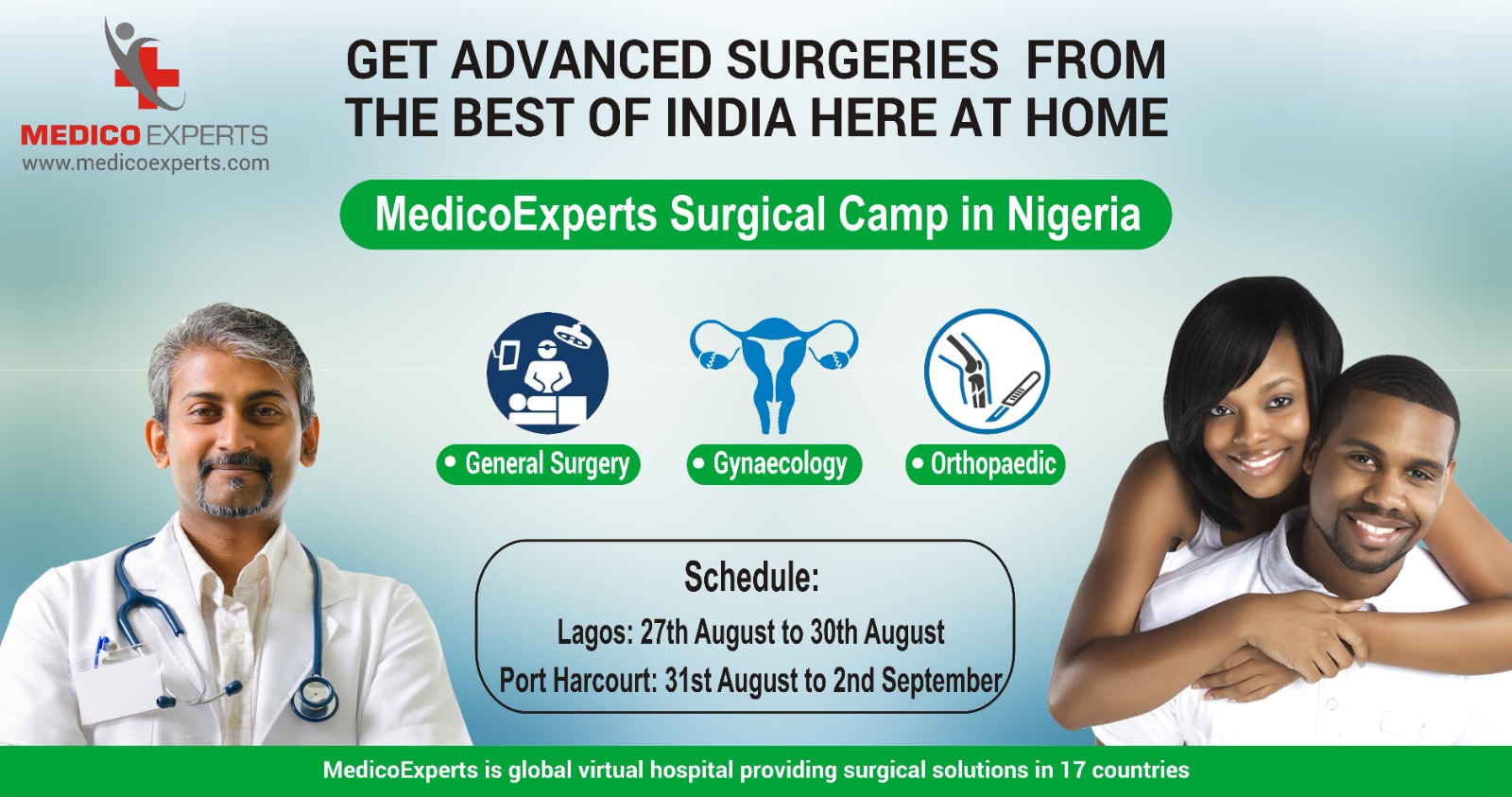
I found MedicoExperts on Social Media – Facebook and they helped me to get the very good treatment done in Nigeria by the best Indian orthopaedic surgeon. Initially, I was scared and did not believe it was possible to get such good treatment in Ibadan, Nigeria where I live, but then I saw the name of the doctor and called MedicoExperts and I was told to visit the MedicoExperts Surgical Centre and get treatment in Nigeria. This has saved us the trouble and cost of travelling and accommodation. I gave this a try and my mother can walk now, and I really impressed with the outcome. I will fully recommend this to anyone out there.

Author Bio
Dr. Subhamoy Mukherjee is a molecular oncologist with experience working with genomic profiles. He has several years of experience in scientific writing. He takes a strong interest in making people aware of different treatment approaches in cancer, and acute and chronic diseases. He also has an interest in innovative approaches for treating different mental and physical illnesses. View all posts by Dr. Subhamoy Mukherjee
Clinically and Medically Reviewed By MedicoExperts Editorial & Clinically Review Board
- https://pmc.ncbi.nlm.nih.gov/articles/PMC4436478/
- https://www.mayoclinic.org/diseases-conditions/acl-injury/symptoms-causes/syc-20350738
- https://www.radiologyinfo.org/en/info/boneradhttps://www.mayoclinic.org/tests-procedures/mri/about/pac-20384768
Medical Disclaimer: The information provided is for general understanding and educational purposes only. It is not intended to replace professional medical advice, diagnosis, or treatment. Always consult your doctor or a qualified healthcare provider for personalized medical guidance.